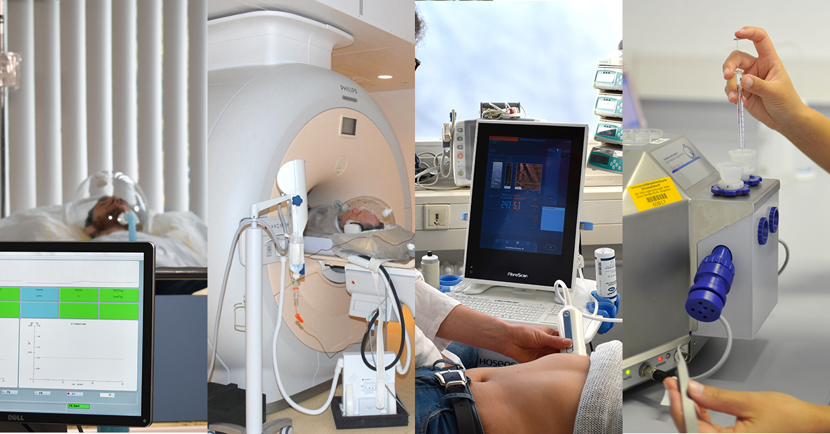
Metabolic phenotyping
We are using the current gold-standard in vivo methodology for assessing pancreatic ß-cell function, tissue-specific insulin sensitivity and nutrient tolerance, e. g. hyperglycemic and hyperinsulinemic-normoglycemic clamps, mixed meal and oral glucose tolerance tests with/without biopsies of skeletal muscle and adipose tissue.
The hyperinsulinemic-euglycemic clamp
is the gold standard method for the assessment of tissue-speciifc insulin sensitivity in vivo, when combined with stable isotope labelled-glucose tracers.
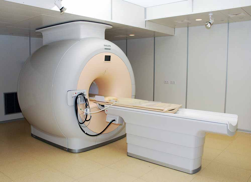
Magnetic resonance imaging (MRI), spectroscopy (MRS) and elastography (MRE)
are non-invasive techniques allowing for examining the body´s organs regarding their morphology (MRI) and biochemical features (MRS). MRI provides 2- or 3-D images of the human body and is used for localization and measurement of organ volume (e. g. liver) and tissue distribution (e. g. fat compartments). MRS makes use of radio waves and the magnetic properties of different metabolites, which allow for exact quantification of their concentrations in tissues. Multinuclear non-invasive in vivo 31P/13C/1H MRS makes it possible to monitor non-invasively the time course of several key metabolites (e. g. liver ATP and triglycerides, muscle glucose-6-phosphate and glycogen) and to assess metabolic fluxes.
MRE makes use of a vibration source to generate low frequency mechanical waves within tissues, imaging of the propagating waves using a phase-contrast MRI. This method allows for quantitative assessment of tissue stiffness and represents a novel tool for non-invasive diagnosing and staging of liver fibrosis.
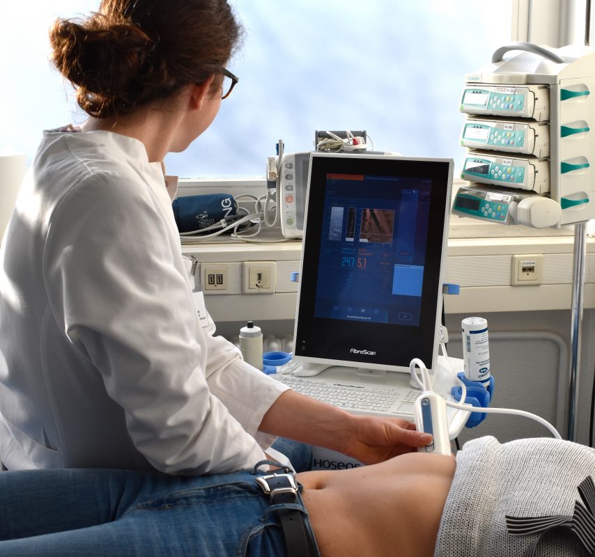
Transient elastography (TE)
is a fast, non-invasive, ultrasound-based technique for measuring liver steatosis and fibrosis. Assessment of steatosis is derived from the controlled attenuation parameter (CAP) (Fibroscan, Echosens). Liver stiffness is evaluated by measuring the velocity of a vibration wave (‘shear wave’) generated on the skin.
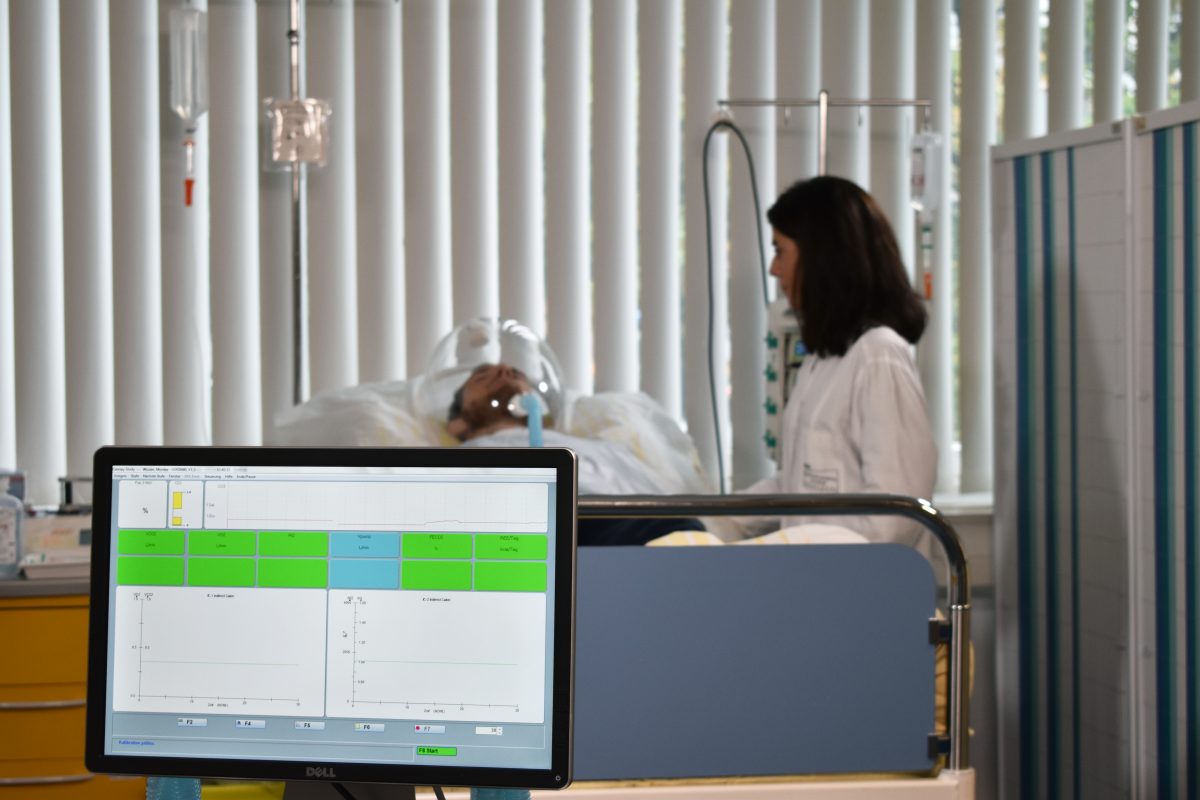
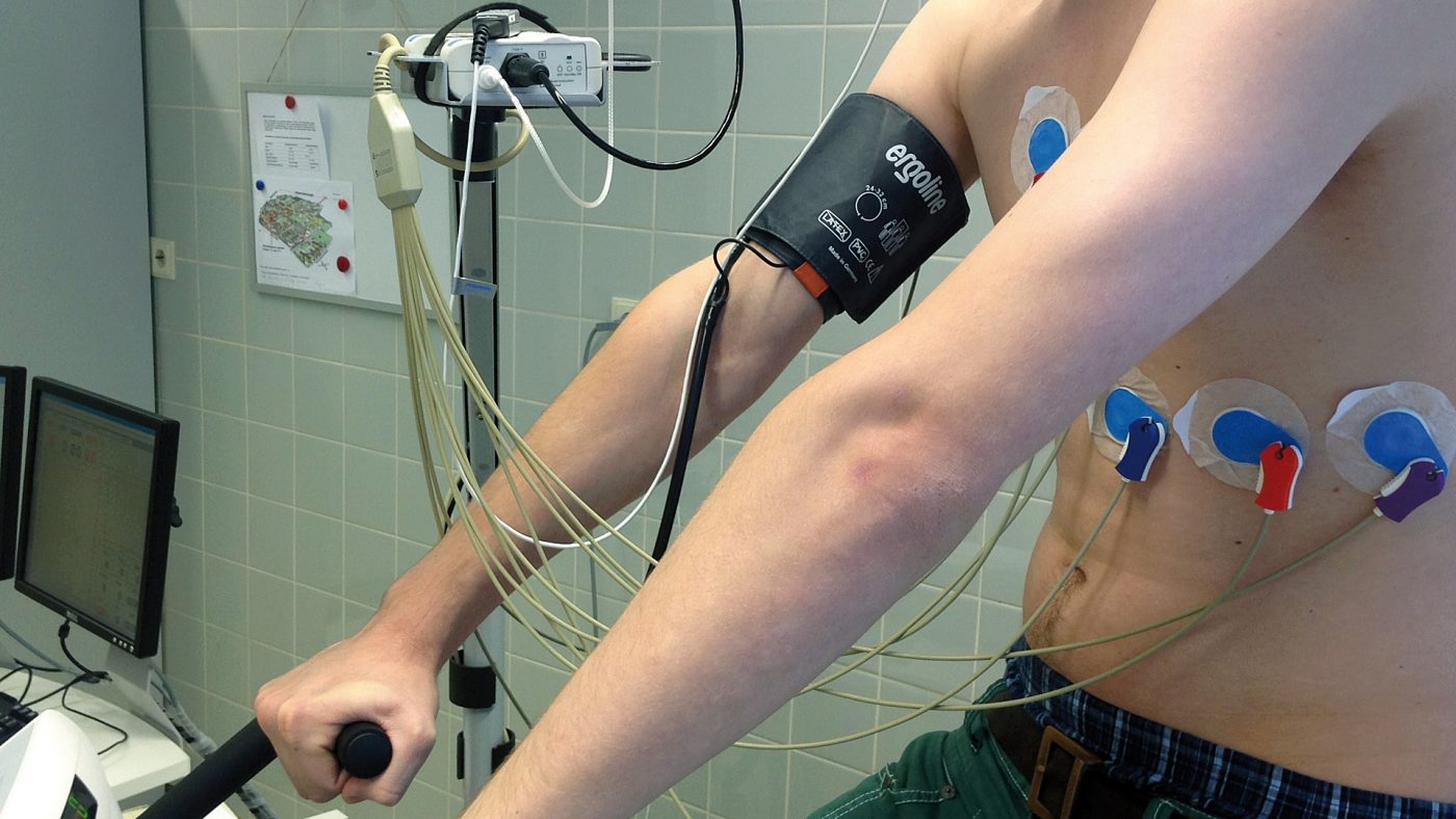
In vivo assessment of energy metabolism
comprises indirect calorimetry (IC) and cycling-spiroergometry or cardiopulmonary exercise testing (CPET). IC is a non-invasive respiratory test measuring carbon dioxide (CO2) production and oxygen (O2) consumption under steady-state conditions. This method yields whole-body energy expenditure for assessment of the resting metabolic rate, thermic effect of food, substrate utilization and metabolic flexibility. CPET is a well-established non-invasive test, assessing cardiorespiratory performance during submaximal exercising. This test monitors cardiac function from electrocardiogram, heart rate and blood pressure as well as maximal oxygen consumption (VO2max) and ventilation thresholds during increasing workloads, reflecting exercise intensity (Watt).
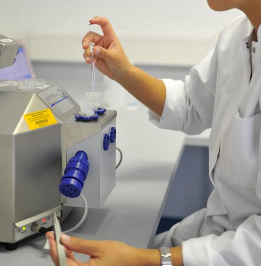
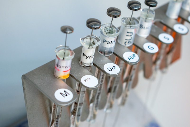
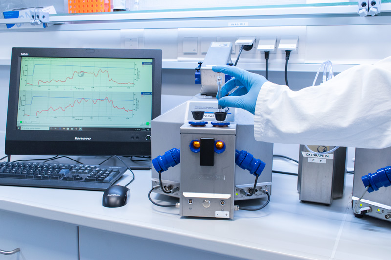
Ex vivo assessment of mitochondrial function
High-resolution respirometry (HRR) is a state-of-the-art approach to assess mitochondrial function in human tissue preparations such as animal tissues and cells. Addition of different substrates, inhibitors and uncouplers allows for detailed analysis of mitochondrial respiratory pathways in the context of metabolic diseases using small amounts of biological samples. This approach can be combined with other modules to assess additional features such as reactive oxygen species (ROS) formation. We are also using other methods for detailed mitochondrial phenotyping including expression of mitochondrial and nuclear genes via polymerase chain reaction (PCR), quantification of complexes of the respiratory system via western blotting, measurement of mitochondrial content via enzymatic assays and transmission electron microscopy from tissue sections as well as assessment of cellular oxidative stress and antioxidant defense systems.
Ex vivo assessment of cell types andextracellular vesicles
Magnetic-activated cell sorting allows for isolating primary cells from tissues using specifically labeled magnetic microbeads. SO, highly pure primary cells populations become available for various analyses. Size exclusion chromatography (SEC) is a rapid and effective method for isolating pure and intact extracellular vesicles (EV), such as exosomes, from biofluids. Indeed SEC columns provide clean samples separated from proteins and lipoproteins, without affecting the structure and function of EV. Nanoparticle Tracking Analysis (NTA) is a real-time technique for assessing count and size of nanoparticles, including EV resuspended in fluid. In addition a fluorescence mode provides differentiation of intrinsic or fluorescently labelled nanoparticles.
In vivo assessment of insulin sensitivity
Hyperinsulinemic-euglycemic clamp is the gold standard method for the assessment of insulin sensitivity in rodents and humans. Under hyperinsulinemic conditions, endogenous glucose production is suppressed. Thus, the glucose infusion rate required to maintain euglycemic concentration of glucose reflects the whole-body insulin sensitivity. Valuable information regarding insulin sensitivity of specific tissues can be obtained upon infusion of labeled-glucose.